Impacted Canines
An impacted tooth simply means that it is “stuck” and cannot erupt into a functional position. Maxillary cuspids, commonly called eyeteeth or canines, are the second most common teeth to become impacted. Cuspids are critical teeth in the dental arch and play an important role in your “bite”. Cuspids are very strong biting teeth and have the longest roots of any human teeth. They are designed to be the first teeth that touch when your jaws close together so they guide the rest of the teeth into the proper bite.
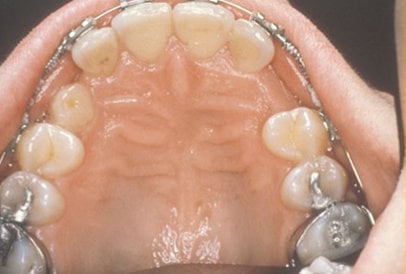
Normally, the maxillary cuspid teeth are the last of the “front” teeth to erupt into place. They usually come in around age 13 and cause any space left between the upper front teeth to close tighter together. If a cuspid gets impacted, every effort is made to get it to erupt into its proper position in the dental arch. The techniques involved to aid eruption can be applied to any impacted tooth in the upper or lower jaw, but most commonly they are applied to the maxillary cuspid teeth. Sixty percent of these impacted eyeteeth are located on the palatal (roof of the mouth) side of the dental arch. The remaining impacted eye teeth are found in the middle of the supporting bone, but are stuck in an elevated position above the roots of the adjacent teeth, or are out to the facial side of the dental arch.
Early Recognition of Impacted Canines is the Key To Successful Treatment
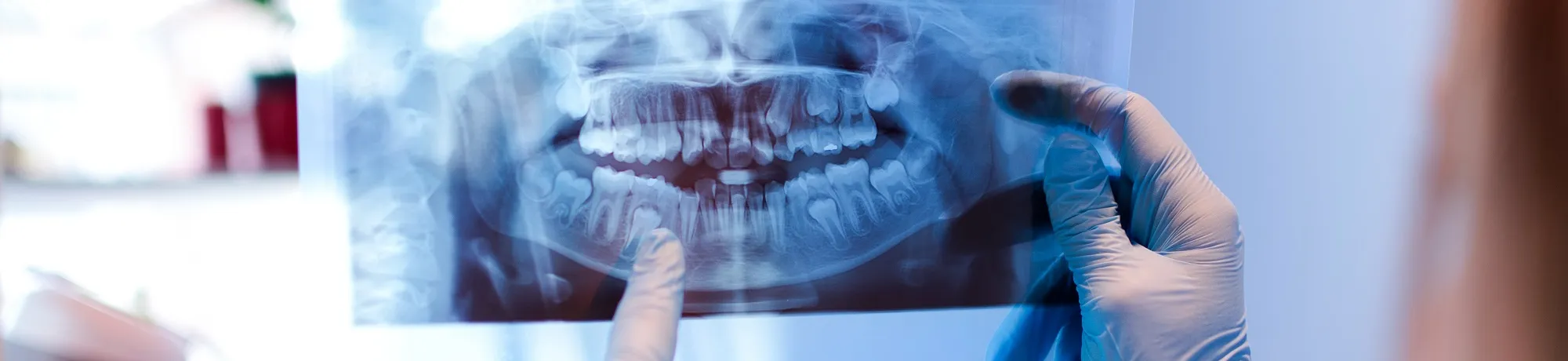
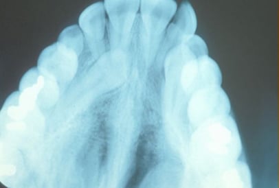
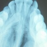
The older the patient the more likely an impacted eyetooth will not erupt by natural forces alone, even if the space is available for the tooth to fit in the dental arch. The American Association of Orthodontists recommends that a panoramic x-ray, along with a dental examination, be performed on all dental patients at the age of seven to count the teeth and determine if there are problems with eruption of the adult teeth. It is important to determine whether all the adult teeth are present or if some adult teeth are missing. Occasionally, your surgeon may need an additional x-ray, called a cone beam CT (CBCT) scan to see a 3-D view of the impacted toot and adjacent teeth.
This exam is usually performed by your general dentist or hygienist who will refer you to an orthodontist if a problem is identified. Treating such a problem may involve an orthodontist placing braces to open spaces allowing for proper eruption of the adult teeth. Treatment may also require referral to an oral surgeon for extraction of over-retained baby teeth and/or selected adult teeth that are blocking the eruption of the all-important eyeteeth. The oral surgeon will also need to remove any supernumerary (extra) teeth or growths that are blocking the eruption of any adult teeth.
If the eruption path is cleared and the space is opened up by age 11 to 12, there is a good chance that the impacted canine will erupt with nature’s help. If the canine tooth is allowed to develop too much under the surface (by age 13-14), the impacted tooth will not erupt by itself, even with the space cleared for its eruption. If the patient is older (over 40), there is a much higher chance that the tooth will be fused in position. In these cases, the tooth will not budge despite all the efforts of the orthodontist and oral surgeon to erupt it into place. Sadly, the only option at this point is to extract the impacted tooth and consider an alternate treatment to replace it in the dental arch (crown on a dental implant or a fixed bridge.)
What Happens if the Canine Will Not Erupt When Proper Space is Available?
In cases where the canines will not erupt spontaneously, the orthodontist and oral surgeon will work together to get these teeth to erupt. Each case must be evaluated on an individual basis, but treatment will usually involve a combined effort between the orthodontist and the oral surgeon. The most common scenario will call for the orthodontist to place braces on the teeth (at least the upper arch.)
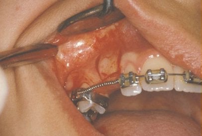
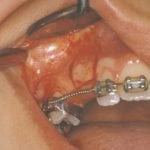
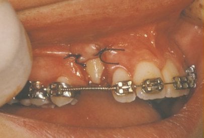
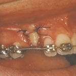
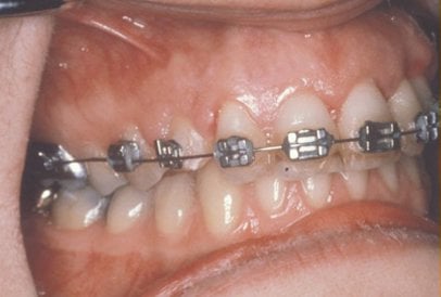
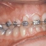
In a simple surgical procedure performed in the surgeon’s office, the gum on top of the impacted tooth will be lifted up to expose the hidden tooth underneath. If there is a baby tooth present it will be removed at the same time. Once the tooth is exposed, the oral surgeon will bond an orthodontic bracket to the exposed tooth. The bracket will have a miniature chain or other attachment attached to it. The oral surgeon will guide the chain back to the orthodontic arch wire where it will be temporarily attached. Often, the surgeon will leave the exposed and impacted tooth uncovered by suturing the gum up high above the tooth, or making a window in the gum covering the tooth.
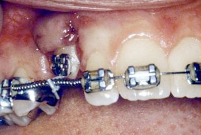
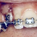
After surgery, the patient will return to the orthodontist. When appropriate, a rubber band will be attached to the chain to put a light eruptive pulling force on the impacted tooth. This will begin the process of moving the tooth into its proper place in the dental arch. This is a carefully controlled, slow process that may take up to a full year to complete. Remember, the goal is to erupt the impacted tooth and not to extract it. Once the tooth has moved into the arch in its final position, the gum around it will be evaluated to make sure it is sufficiently strong and healthy to last for a lifetime of chewing and tooth brushing.
Exposure and Bracketing of an Impacted Cuspid
These basic principals can be adapted to apply to any impacted tooth in the mouth. It is not that uncommon for both of the maxillary cuspids to be impacted. When the orthodontist is ready, the surgeon will expose and bracket both teeth in the same visit so that the patient only has to heal from one surgery. Because the anterior teeth (incisors and cuspids) and the bicuspid teeth are small and have single roots they are easier to erupt if they get impacted than the posterior molar teeth. The molar teeth are much bigger teeth and have multiple roots making them more difficult to move. The orthodontic maneuvers needed to manipulate an impacted molar tooth can be more complicated because of their location in the back of the dental arch.
Recent studies have revealed that with early identification of impacted eyeteeth (or any other impacted tooth other than the wisdom teeth,) treatment should be initiated at a younger age. Once the general dentist or hygienist identifies a potential eruption problem, the patient should be referred to the orthodontist for early evaluation. In some cases, the patient will be sent to the oral surgeon before braces are even applied to the teeth. As mentioned earlier, the surgeon will be asked to remove over-retained baby teeth and/or selected adult teeth. He will also remove any extra teeth or growths that are blocking the eruption of the developing adult teeth. Finally, he may be asked to simply expose an impacted eyetooth without attaching a bracket and chain to it. In reality, this is an easier surgical procedure to perform than having to expose and bracket the impacted tooth. This will encourage some eruption to occur before the tooth becomes totally impacted (stuck). By the time the patient is at the proper age for the orthodontist to apply braces to the dental arch, the eyetooth will have erupted enough so that the orthodontist can bond a bracket to it and move it into place without needing to force its eruption. This saves time for the patient and means less time in braces – always a plus for any patient!
What to Expect from Surgery to Expose & Bracket an Impacted Tooth
The surgery to expose and bracket an impacted tooth is a very straightforward surgical procedure that is performed in the oral surgeon’s office. For most patients, it is performed using laughing gas and a local anesthetic. In selected cases, it will be performed under IV sedation if the patient desires to be asleep. These issues will be discussed in detail at your preoperative consultation with your doctor. (You can also refer to Preoperative Instructions under Surgical Instructions on this website for a review of any details.)
You can expect a limited amount of bleeding from the surgical site after surgery. A soft, bland diet is recommended at first, but you may resume your normal diet as soon as you feel comfortable chewing. It is advised that you avoid sharp food items, such as crackers and chips, as they will irritate the surgical site if they jab the wound during initial healing. Often, the surgical site may be covered by a dressing, which resembles pink putty. This dressing generally remains in place for a week and will be removed by your surgeon at a follow-up visit. Sometimes, this dressing will fall out before the follow-up visit. If this happens, simply call our office on the next business day. Rarely does this dressing need to be replaced. Your doctor will see you seven to ten days after surgery to evaluate the healing process and make sure you are maintaining good oral hygiene. As always, your doctor is available at the office and can be contacted after hours if any problems should arise after surgery. Simply call Kalamazoo Oral & Maxillofacial Surgery at Portage Phone Number 269-323-1527 if you have any questions.